

Abstract
Introduction
Blood coagulation factor XIII (FXIII) is a precursor of the active transglutaminase (FXIIIa) that catalyzes covalent cross-linking within polymeric fibrin to increase mechanical and proteolytic stability of blood clots and thrombi. Structurally, inactive plasma FXIII (pFXIII) with a molecular mass of 326 kDa is a heterotetramer (A2B2) with two catalytic A subunits (FXIII-A, 83 kDa) and two non-catalytic B subunits (FXIII-B, 80 kDa).
The inactive pFXIII is converted into a catalytically active form by thrombin and Ca2+. Thrombin cleaves off an activation peptide (AP) from the N-terminus of each A subunit, and subsequently B subunits dissociate from thrombin-cleaved A subunits in presence of Ca2+. However, the oligomerization state of activated A and B subunits remain controversial.
Both activated and inactive FXIII A subunits have been characterized crystallographically, while the atomic structure of the FXIII-A2B2 complex remains unknown. Therefore, our goal was to visualize and characterize the molecular structure of FXIII and its activated form FXIIIa using high-resolution atomic force microscopy (AFM).
Methods
Commercially available pFXIII (Enzyme Research Laboratories, USA), non-activated or activated with thrombin and Ca2+, was adsorbed on the surface of graphite rendered hydrophilic with an amphiphilic hydrocarbon-glycine modifier followed by single-molecule AFM imaging, methodology developed by us to allow high-resolution imaging. Additionally, recombinant rFXIII-A2 (Zymogenetics, USA) and recombinant rFXIII-B2 (Zedira, Germany) were imaged to compare the structures and dimensions of FXIII and FXIIIa with individual dimeric subunits.
Results
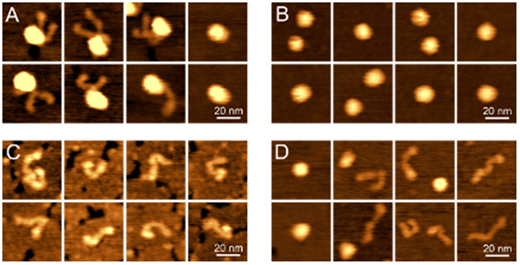
Individual molecules of pFXIII were visualized in AFM as globules with two (67% of the molecules), one (14%), or no (19%) thread-like extensions (Fig. 1A). The size (height) of the globular portions had a symmetrical distribution with a maximum at 3.5 nm. The strands had a peak contour length of 20 nm and a height of 0.4 nm. Based on the known polypeptide composition of pFXIII-A2B2, the observed molecular images could be interpreted as an A2 globule with one or two individual B subunits extending out from it. The structural diversity of FXIII forms may reflect an equilibrium between association and dissociation of B subunits from the A2 dimer. Alternatively, the B subunits can be partially or fully wrapped around the globular A2 dimer, making them invisible in AFM images.
Although a crystallographic structure of FXIII-A2 is available, its AFM imaging was important to identify the A2 dimer within inactive and activated pFXIII molecules: rFXIII-A2 was a globule with the same height as the pFXIII globular core (Fig. 1B).
Recombinant dimeric B subunits (rFXIII-B2) appear as thin flexible strands with an average contour length of 33 nm and a height of 0.6 nm (Fig. 1C). Thus, both the length and height of the free B2 dimers were significantly larger compared to the strands sticking out from the globular core of pFXIII-A2B2 molecules, we conclude that these strands in pFXIII-A2B2 comprise monomeric B subunits.
In thrombin-activated pFXIIIa, the B-subunits dissociated from the A2 dimer and self-associated to form B2 dimers (Fig. 1D). The height distribution of globular particles remaining separated was bimodal with two peaks: one at 3.3 nm corresponded to the whole tetrameric pFXIII-A2B2 and, potentially, dimeric FXIII-A2, while the second major peak at 2.1 nm likely represented FXIII-A monomers.
Conclusions
Our results provide qualitative and quantitative molecular structural characteristics of inactive and activated human pFXIII as well as dimeric A and B subunits. We have shown that inactive plasma FXIII (pFXIII-A2B2) consists of a globular A2 dimer core and individual B subunits extending out as flexible strands. Recombinant inactive FXIII-A subunits in the absence of FXIII-B subunits form a globular homodimer. Recombinant FXIII-B subunits in the absence of FXIII-A subunits form flexible homodimeric strands. Upon activation of pFXIII-A2B2 with thrombin and Ca2+, the initially monomeric B subunits dissociate from the globular core of the molecule and form B2 homodimers, while the initially dimeric activated A subunits dissociate into monomers.
Fig. 1. AFM images of A: pFXIII-A2B2, B: rFXIII-A2, C: rFXIII-B2, D: thrombin-activated pFXIIIa.
No relevant conflicts of interest to declare.

This feature is available to Subscribers Only
Sign In or Create an Account Close Modal